Retinal Vein Occlusion: Risk Factors and Injections Explained
 Dec, 24 2025
Imagine waking up one morning and noticing your vision is blurry - not like you’re tired, but like someone smeared grease across your glasses. No pain. No warning. Just sudden, silent vision loss in one eye. That’s what retinal vein occlusion (RVO) often feels like. It’s not rare. Around 16.4 million people worldwide live with it. And for many, it’s not just a one-time event - it’s the start of a long road of treatments, injections, and constant monitoring.
What Exactly Is Retinal Vein Occlusion?
Your retina is the light-sensitive layer at the back of your eye. It turns images into signals your brain understands. Blood flows into the retina through arteries and leaves through veins. When one of those veins gets blocked - usually because of a clot or pressure from a hardened artery - blood backs up. Fluid leaks into the retina, swelling the macula (the part you use for sharp central vision). That’s RVO. There are two main types:- Central Retinal Vein Occlusion (CRVO): The main vein is blocked. Vision loss tends to be more severe.
- Branch Retinal Vein Occlusion (BRVO): Only a smaller branch is blocked. Vision loss is often partial - maybe just the top or bottom half of your vision.
Who’s Most at Risk?
RVO doesn’t pick favorites, but it does favor certain people. Over 90% of CRVO cases happen in people over 55. Half of all cases occur in those 65 and older. But it’s not just an older person’s disease - 5 to 10% of cases strike people under 45. Here’s what increases your risk the most:- High blood pressure: Present in up to 73% of CRVO patients over 50. It’s the #1 risk factor. Uncontrolled hypertension makes blood vessels stiff and prone to clotting.
- High cholesterol: About 35% of RVO patients have total cholesterol above 6.5 mmol/L. Fatty deposits narrow blood vessels, making blockages more likely.
- Diabetes: Affects about 10% of RVO patients over 50. It damages blood vessel walls over time, making them leaky and fragile.
- Glaucoma: High pressure inside the eye can physically compress the retinal vein, especially where it passes through the optic nerve.
- Smoking: Found in 25-30% of cases. It thickens blood, damages vessel linings, and speeds up artery hardening.
- Obesity and inactivity: Both raise inflammation and blood pressure, indirectly raising RVO risk.
How Do Doctors Diagnose It?
It starts with a simple eye exam. Your doctor will check your vision and look at the back of your eye with a special lens. But the real clues come from advanced imaging:- Optical Coherence Tomography (OCT): This scan shows the thickness of your retina. If the macula is swollen (macular edema), it shows up clearly - usually when central subfield thickness exceeds 300 micrometers.
- Fluorescein angiography: A dye is injected into your arm, and a camera tracks how it flows through the retinal vessels. It shows exactly where the blockage is and if new, leaky blood vessels are forming.
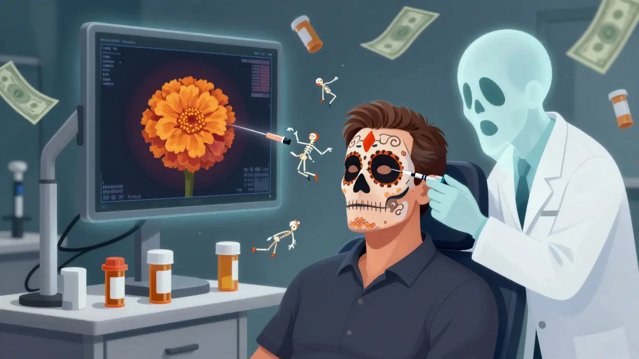
The Role of Injections: Anti-VEGF and Steroids
There’s no magic cure to unblock the vein. Treatment focuses on stopping the damage - mainly the swelling and abnormal blood vessel growth. The go-to treatment? Anti-VEGF injections. VEGF is a protein that causes leaky blood vessels and swelling. Blocking it reduces fluid buildup and can restore vision. Three drugs are used:- Ranibizumab (Lucentis): Approved for RVO in 2010. Clinical trials showed patients gained an average of 16.6 letters of vision in a year.
- Aflibercept (Eylea): Approved in 2012. In trials, patients gained 18.3 letters on average. Studies now show it works better than others when baseline vision is worse than 20/50.
- Bevacizumab (Avastin): Originally a cancer drug, it’s used off-label because it’s cheap - about $50 per shot vs. $2,000 for the others. It works just as well for many patients.
- Red spot on the white of the eye (subconjunctival hemorrhage) - happens in 25-30% of cases
- Temporary increase in eye pressure - 15-20% of patients
- Floaters - 10% of cases
- Endophthalmitis (serious eye infection) - less than 0.1% of injections
What About Steroid Injections?
If anti-VEGF doesn’t work well enough, doctors turn to steroids. The dexamethasone implant (Ozurdex) is a tiny, dissolvable pellet injected into the eye. It slowly releases steroid over 3-6 months. In the GENEVA trial, 27.7% of CRVO patients gained 15 or more letters of vision with Ozurdex - compared to 12.9% with placebo. That’s a big jump. But steroids come with trade-offs:- Up to 70% of patients develop cataracts faster
- 30% get elevated eye pressure, sometimes needing medication or surgery
How Often Do You Need Injections?
It’s not a one-and-done fix. Most patients need monthly shots for 3-6 months until swelling clears. Then, doctors switch to “as needed” dosing - checking with OCT every 4-8 weeks. Real-world data shows patients get 8-12 injections per year on average. The goal? Keep central subfield thickness below 250 micrometers. Newer protocols like treat-and-extend are changing the game. Instead of fixed monthly shots, doctors gradually stretch the time between injections if the eye stays stable. One 2023 study found this approach cut injection frequency by 30% without losing vision gains.What’s the Real-World Experience Like?
Patients don’t just see numbers on a chart. They feel the cost, the fear, the fatigue. One man, 62, started monthly Lucentis shots after CRVO. His vision improved from 20/200 to 20/60 - life-changing. But each injection cost $150 out-of-pocket. On a fixed income, that added up fast. Another patient tried eight Avastin shots with little improvement. Then she got the Ozurdex implant. Ten lines of vision gained. Worth the $2,500 price tag, she says. But the emotional toll is real. People talk about anxiety before each shot. Some develop “injection fatigue” - missing appointments because the stress becomes too much. One patient stopped going after 18 months, even though her vision kept improving. A 2022 survey of over 1,200 RVO patients found:- 78% saw significant vision improvement after a year of anti-VEGF therapy
- 63% struggled with financial burden
- 41% felt overwhelmed by treatment frequency

What’s Next for RVO Treatment?
The future is about reducing burden. Researchers are testing:- Extended-delivery systems: Like Susvimo - a tiny pump implanted in the eye that releases ranibizumab for months at a time. It’s approved for AMD and in trials for RVO.
- Gene therapy: RGX-314 delivers a gene that makes your eye produce its own anti-VEGF protein. Early trials show promise for long-term suppression without injections.
- New drugs: OPT-302 blocks a different VEGF protein (VEGF-C/D) and is being tested alongside aflibercept for stubborn cases.
Can You Prevent RVO?
You can’t undo aging, but you can control your risks:- Keep blood pressure under 130/80
- Manage cholesterol with diet, exercise, and meds if needed
- Control blood sugar if you have diabetes
- Quit smoking - it’s the single biggest lifestyle change you can make
- Exercise regularly - even a 30-minute walk daily helps circulation
- Get annual eye exams - especially if you’re over 50 or have risk factors
Final Thoughts
Retinal vein occlusion isn’t just an eye problem - it’s a warning sign. It’s your body telling you something’s wrong with your blood vessels. And while injections can restore vision, they’re not a cure. The real win is preventing it in the first place. If you’ve been diagnosed, stick with your treatment. Don’t skip appointments. Ask about cost-saving options like bevacizumab. Talk to your doctor about treat-and-extend plans. And remember - vision improvement is possible. Many people go from barely seeing to reading again. It takes time, patience, and persistence.Can retinal vein occlusion be cured?
No, RVO cannot be cured. The blocked vein doesn’t reopen on its own. But treatments like anti-VEGF injections and steroids can stop the swelling and fluid buildup that cause vision loss. Many patients regain significant vision - some even to 20/40 or better - with consistent treatment. The goal is managing complications, not reversing the blockage.
Are RVO injections painful?
Most patients feel only slight pressure or a brief sting. The eye is numbed with drops before the injection. The needle is very thin, and the procedure takes less than 10 minutes. The anxiety before the shot is often worse than the procedure itself. Serious pain is rare.
How long do the effects of anti-VEGF injections last?
Each injection typically works for 4 to 6 weeks. That’s why monthly dosing is common at first. As the swelling improves, doctors may extend the time between shots - sometimes to every 8 or 12 weeks - if the eye stays stable. The goal is to use the fewest injections needed to maintain vision.
Can I drive after an RVO injection?
Not right away. Your pupil will be dilated, and your vision may be blurry for several hours. Most clinics recommend you have someone drive you home. Avoid driving or operating heavy machinery until your vision clears, usually within a few hours.
Is Avastin safe for RVO if it’s not FDA-approved for this use?
Yes. Avastin (bevacizumab) is used off-label for RVO, but it’s one of the most studied and safest options. Multiple large studies show it works just as well as FDA-approved drugs like Lucentis and Eylea. It’s used in over 60% of injections at safety-net clinics because it’s affordable and effective. The injection procedure and risks are identical to the branded drugs.
What happens if I skip my RVO injections?
Skipping injections can lead to worsening vision. Fluid builds up again, causing more damage to the retina. In some cases, abnormal blood vessels grow and bleed inside the eye, causing sudden, severe vision loss. Even if your vision seems stable, ongoing treatment is needed to prevent relapse. Consistency is key.
Can RVO affect both eyes?
It’s possible, but uncommon. Most people experience RVO in one eye first. However, if you have systemic risk factors like high blood pressure, diabetes, or clotting disorders, your other eye is at higher risk. Regular eye exams are critical to catch early signs in the second eye.
Is there a link between RVO and stroke or heart disease?
Yes. RVO is a sign of widespread vascular disease. People with RVO have a higher risk of stroke, heart attack, and other vascular events in the next 5 years. That’s why doctors often refer RVO patients to their primary care provider or cardiologist for full cardiovascular screening - blood pressure, cholesterol, glucose, and clotting tests.
Oluwatosin Ayodele
December 26, 2025 AT 07:31Let me cut through the BS-this whole anti-VEGF industry is a cash grab. Avastin works just as good as Lucentis, but they charge 40x more because the FDA rubber-stamped it for eyes. Big Pharma doesn’t care if you go blind-they care if you keep coming back for monthly shots. I’ve seen patients drain their retirement funds just to keep seeing their grandkids. The system is rigged.
Jason Jasper
December 26, 2025 AT 14:25I appreciate the breakdown, especially the stats on cholesterol and hypertension. I had BRVO last year-no symptoms until I couldn’t read my phone. Turns out my BP was 168/102 and I didn’t even know it. Since then, I’ve lost 30 lbs, quit soda, and started walking daily. My OCT scan improved by 80 microns in 6 months. Prevention isn’t sexy, but it’s the only real win.
Mussin Machhour
December 27, 2025 AT 18:18Y’all need to stop treating this like it’s just an eye thing. RVO is your body screaming that your blood is basically sludge. I got CRVO at 52, started on Avastin, and now I’m on Ozurdex because my doc said I was ‘injection-fatigued.’ Honestly? I’m just glad I can still see my dog’s face. But man, the anxiety before each shot? Like waiting for a dentist drill. You’re not just fighting fluid-you’re fighting fear every time.
Winni Victor
December 28, 2025 AT 13:16So let me get this straight-some guy in a lab invented a protein blocker, then sold it for $2000 a shot, but the same damn thing costs $50 if you use the cancer version? And we’re supposed to be grateful? The only thing ‘miraculous’ here is how many people still believe the system isn’t broken. I’d rather go blind than fund another billionaire’s yacht.
Christopher King
December 30, 2025 AT 08:35They don’t tell you this, but RVO isn’t a disease-it’s a cover-up. The real cause? 5G towers frying the retinal capillaries. Look at the data-cases exploded right after cell towers went nationwide. And why are they pushing injections? Because they want you dependent. The implant? It’s got a microchip. I read the patent. It tracks your eye movements. They’re not fixing your vision-they’re monitoring you. Wake up.
Zabihullah Saleh
December 30, 2025 AT 11:02Back home in Afghanistan, my uncle had this. No fancy OCT, no injections-just a village doctor with a magnifying glass and some herbal drops. He lost vision in one eye, but kept the other. We didn’t have drugs, but we had patience. I think we lost something modern medicine can’t replace: time. We sat. We waited. We prayed. Now we rush to fix it with needles and bills. Maybe we’re treating the symptom and forgetting the soul.
Linda B.
December 31, 2025 AT 19:18While I appreciate the clinical detail presented, I must note that the omission of any reference to the potential iatrogenic effects of repeated intraocular injections-particularly the cumulative endothelial cell loss and retinal toxicity-is not merely an oversight, but a glaring ethical lacuna in contemporary ophthalmic discourse. The normalization of this invasive paradigm, absent long-term longitudinal data, constitutes a form of medical hubris masquerading as progress.
Carlos Narvaez
January 1, 2026 AT 10:35Avastin works. Period. Stop overpaying. Also, if you’re over 50 and haven’t had an eye exam in 2 years, you’re playing Russian roulette with your vision. Your optometrist isn’t just selling drops-they’re saving your life.